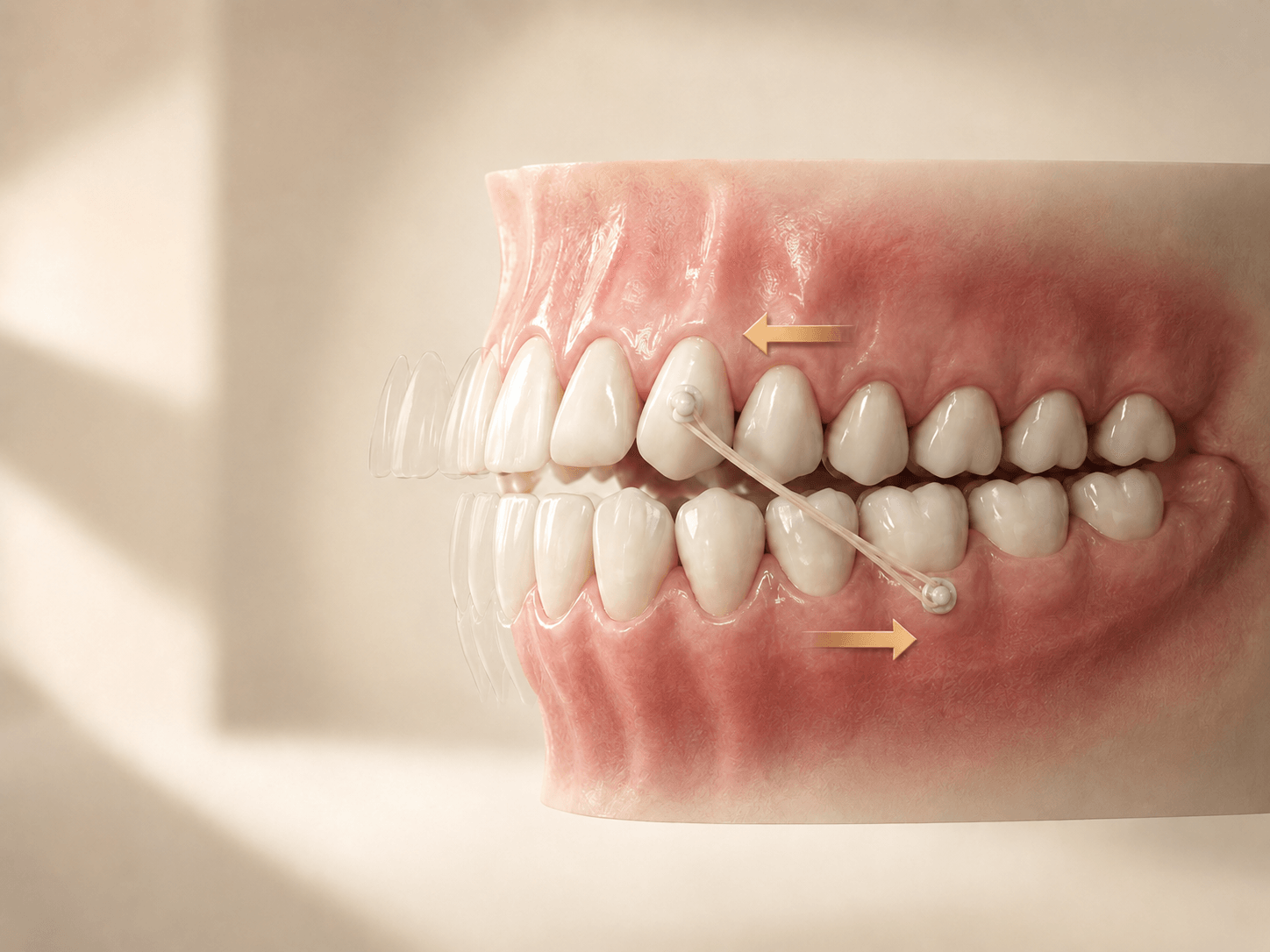
Risk assessment & planning
Review complexity, movement predictability, staging, expected duration, compliance demands and package scope before the first aligner is approved.
Review pre-treatment support
LUMOSCO connects pre-treatment risk review, structured planning guidance and in-treatment monitoring in one doctor-led workflow.
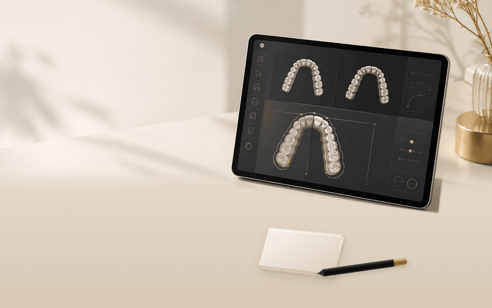
The LUMOSCO design team organizes case observations into two linked phases, helping the practice understand risk before approval and monitor how the case is tracking after delivery.
Review complexity, movement predictability, staging, expected duration, compliance demands and package scope before the first aligner is approved.
Review pre-treatment supportUse fit checks, updated scans and plan comparison to support timely recommendations, next-set ordering and refinement decisions.
Review monitoring supportRepresentative reviews turn technical observations into practical points the doctor can evaluate and discuss with the patient before treatment begins.
Instead of generic comments, the service points to the movement, contact or design decision that needs the doctor’s attention.
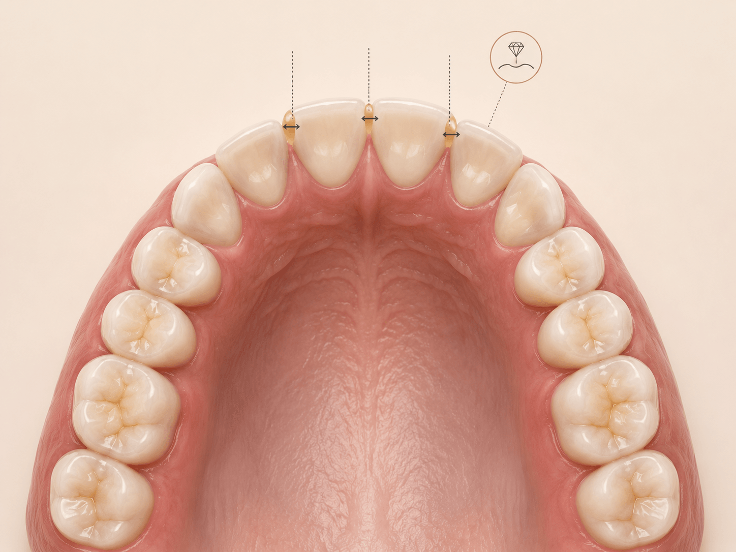
Confirm sufficient space at every annotated contact. The redrawn figure restores the source values around the arch—including 0.16, 0.24, −0.11, 0.11, −0.05 and 0.20—together with the posterior contact and 0.5 movement markers. When space is inadequate, perform additional interproximal reduction as needed to create room for the upcoming movement.
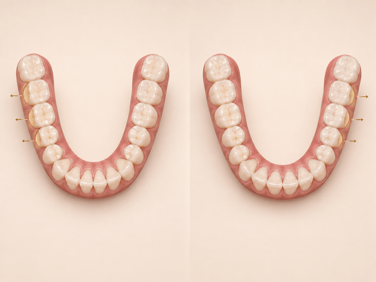
For this staged example, order 12 trays for the first set, check fit at tray 9 and submit an updated scan. If everything fits and tracks well, release the following trays. The original setup also compares distalization and IPR annotations at stages 6 and 15.
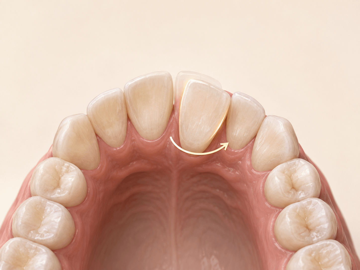
Flag the notable mesial rotation of tooth #9. The source note indicates that this movement will probably be corrected during the final refinement phase, allowing the doctor to plan the pathway and communicate it early.
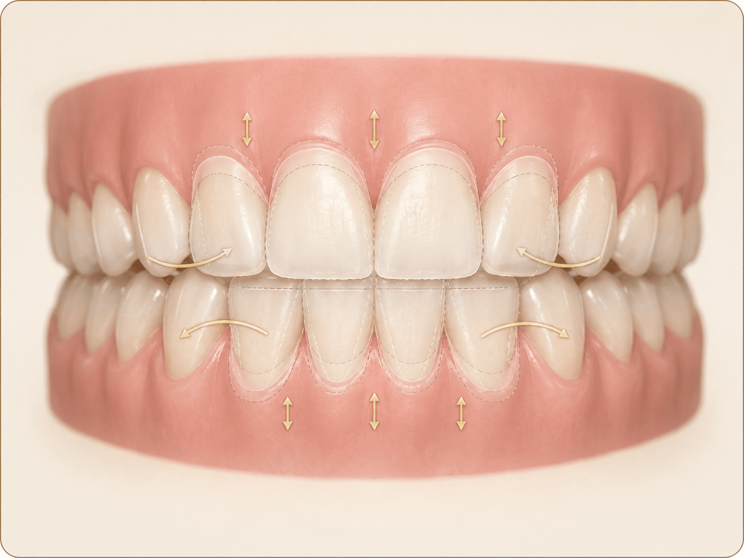
Use further digital overcorrection for the upper and lower anterior teeth in both the vertical dimension and labiolingual torque control. Clinically, final occlusal opening is anticipated to reach about 70% of the opening shown by the red line in the animation.
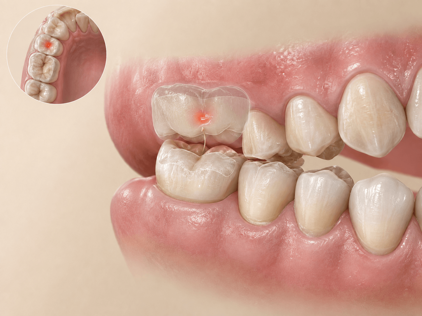
In the source case, habitual right-sided chewing is associated with abnormal occlusal wear and an early contact on tooth #2. Intentional overcorrection of #2 is designed to minimize occlusal interference and further wear.
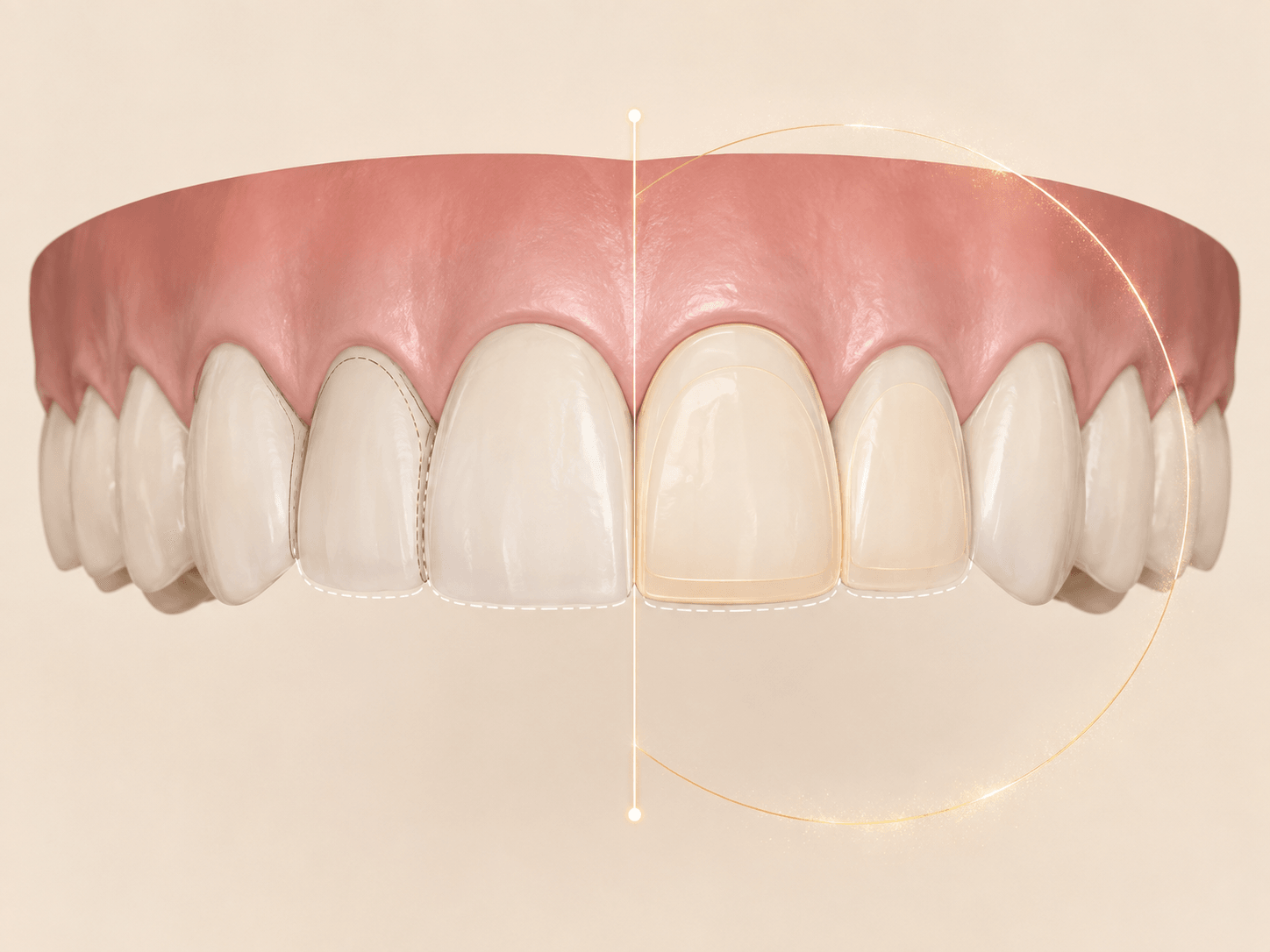
The source plan virtually removes existing bonding and optimizes alignment with relapse control in mind. For maxillary anterior symmetry, compare restorative modification of #9 and #10 with enamel reduction of #7 and #8; the stated preference is restorative modification.
Representative, de-identified planning examples. They illustrate communication and workflow support—not a substitute for the treating doctor’s diagnosis, prescription or clinical judgment.
When the patient has about two aligners left, the practice can capture the latest clinical position and use it to inform the next treatment step.
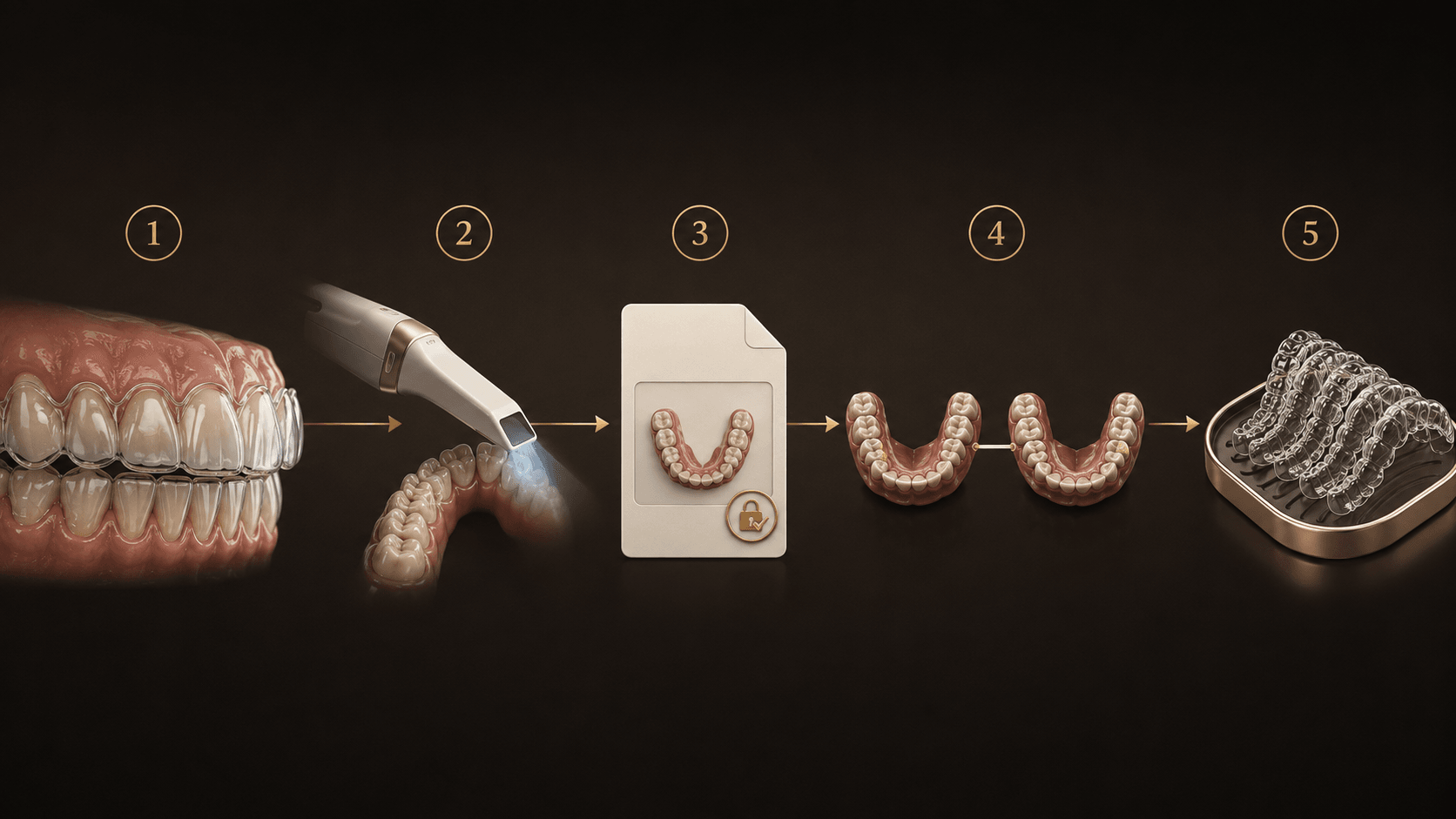
When approximately two aligners remain in the current set, evaluate seating, tracking and patient wear.
Capture an updated intraoral scan so the current tooth positions can be reviewed accurately.
Add the scan and a concise clinical note to the follow-up record for design-team review.
Compare the latest scan with the approved treatment plan and identify any meaningful deviation.
Receive targeted recommendations, confirm the clinical direction and order the next aligner set or refinement.
The treating doctor selects the patient, defines clinical goals, evaluates recommendations and gives final approval at every decision point.
The design team organizes records, highlights risks, prepares reviewable setups and provides technical observations during follow-up.
Start with a case conversation and see how LUMOSCO can support the path from risk review to follow-up.